Category

ATP test

FREE ATP VS INTRACELLULAR ATP

What is ATP?

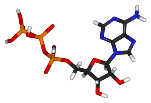
Adenosine triphosphate (ATP) is a molecule used by all living cells to provide energy to metabolic reactions. It is often referred to as the “molecular unit of currency” of intracellular energy transfer. As ATP is specific to living environments, its presence proves the existence of living organisms.
ATP testing is a technique measuring the level of ATP in a sample in just a few minutes.
There are two forms:

  • Free or extracellular ATP,
  • Intracellular ATP.
Molécule ATP
Free ATP

It is the ATP freed by the dying or dead cells. When a cell dies, it loses its membrane integrity. The ATP, which is a very small molecule, is released in the environment.

As it is an unstable molecule in unbuffered water, it is rapidly destroyed. Its stability depends on many parameters:

  • pH: free ATP is stable at neutral pH but its degradation rate quickly increases with alkaline and acidic pH.
  • Temperature: degradation rate increases with temperature. Rather stable at 4°C, it degrades quickly at 25-30°C.
  • Presence of stabilisers: such as polycations and/or some cations.
  • Type of biocide used: oxidising biocides such as chlorine or bromine rapidly degrades free ATP whereas non oxidising biocides have little effect.
  • Presence of other microorganisms: some are able to retrieve free ATP to use it.

Therefore, free ATP stability is difficult to assess because it depends on many parameters. Indeed, if the environment provides a good stability, free ATP will accumulate. However, if the environment is unfavorable, free ATP will disappear very quickly.

Intracellular ATP

It is the ATP found in the living cells. As mentioned above, this molecule plays a vital role in intracellular energy transfer. It is permanently renewed and recycled in the cell, but its production immediately stops when the cell dies.

Total ATP is defined as the sum of free ATP and intracellular ATP. To measure it, it is necessary to destroy the cells with a lysis buffer in order extract the ATP. The measurement is done on the freed intracellular ATP and the free ATP.

To assess the quantity of microorganisms in a sample, only intracellular ATP must be measured.

Total ATP – free ATP = intracellular ATP

A short-sighted and risky approach…

How to measure intracellular ATP?

To quantify intracellular ATP, two different approaches have been developed: an indirect measurement and a direct measurement.

The indirect measurement:

It was the first technique to be developed. It is based on the assumption:

Total ATP – free ATP = intracellular ATP

Intracellular ATP is deduced from the measurement of total and free ATP.

  • Measurement of free ATP: it is measured by bioluminescence without lysis buffer. Thus, only extracellular (free) ATP is able to react with the bioluminescence enzyme. However, as described above, the quantity of free ATP varies greatly depending on many factors. It is not reprensentative of the quantity of microorganisms in the sample. Therefore, the measurement uncertainty is high.
  • Measurement of total ATP: it is measured by bioluminescence with lysis buffer that destroys the cells. Intracellular ATP is freed and add up to the free ATP. As the volumes of sample are generally low (around 100 µl), the presence of biofilm fragments strongly affects the result.

Consequently, using this strategy, the measurement of intracellular ATP is based on the subtraction of two uncertain measurements, which gives an approximate or even a false result.

The other problem lies in the fact that ATP measurement is relative. Indeed, the result is given in Relative Light Unit by the luminometer (RLU). Yet, in this indirect approach, the measurement is not standardised. It depends on many parameters affecting the enzyme efficacy (temperature, effect of lysis buffer, biocides…).

Working on qualitative measurements only leads to important approximations. It is even possible to get higher quantity of free ATP than total ATP!

In conclusion, the measurement of intracellular ATP by this approach is quick and easy. However, given the issues described above, the result can be bias and thus difficult to interpret. Therefore, it is essential to cautiously process the data to avoid over-interpretations.

Direct measurement of intracellular ATP

An extra step is required to measure intracellular ATP directly: filtration on a membrane to eliminate the free ATP. Indeed, as this molecule is very small, it passes through the membrane whereas the microorganisms are retained.

In this approach, the microorganisms retained on the filter are then destroyed in order to free the intracellular ATP. It gives a representative view of the living organisms in the sample.

Besides, this strategy has the advantage of analysing a representative volume of sample (generally between 10 and 50 ml).

Although requiring a little more handling, this approach, combined with standardisation, gives quantitative results much more reliable and comparable in time and space.

Conclusion

ATP testing is one of the fastest and easiest methods to detect microorganisms in water. This 40-year-old analysis has obviously evolved over time.

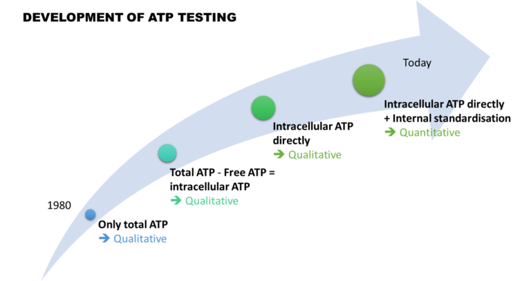
At first, only total ATP was detected qualitatively. Then, thanks to the measurement of free ATP, it was possible to indirectly assess the quantity of intracellular ATP. However, due to high variability, the result interpretation remained complicated.

Then, 15 years ago, new approaches emerged. They now include a filtration step to overcome the problems of variability.

Finally, the integration of external and then internal standardisation made this analysis quantitative. It became robust and comparable in time and space, making ATP testing a relevant analysis.

HOW TO PROPERLY COMPARE ATP TESTING AND CULTURE?

ATP testing and bacterial culture on solid agar plates are two completely different techniques. While culture method only measures culturable bacteria, i.e., the ones able to grow on a given media, ATP testing measures the quantity of ATP in a sample. As this molecule is produced and found in all living bacteria, ATP testing measures all the bacteria, culturable or not. This major difference makes them difficult to compare. 

However, when validating a new technique, it is natural to compare it with the conventional method. To avoid bias in result interpretation, here are some general tips.

General tips, not limited to these two techniques


  • Be aware of what each technique measures: ATP test measures the ATP and thus indirectly the total bacteria, while culture only measures culturable bacteria.
    .
  • Each technology has its own limits. ATP test uses a defined convention to estimate the number of bacteria in the sample (1 pgATP ≈ 1 000 bacteria). As for the culture method, it does not count the VBNC (Viable But Nonculturable) bacteria. Yet, according to literature, only 0.01 to 1% of bacteria grow on HPC media. Bacterial culture is limited by the choice of culture media, of incubation time and incubation temperature.
    .
  • It is necessary to work on a large concentration range, i.e., on several LOG.
    .
  • Perform each measurement at least in triplicate to obtain a significative value for each method.
    .
  • All the samples must be treated in the same way, whatever the analysis method. One of the most frequent mistakes is to collect the sample in a bottle with sodium thiosulfate for culture analysis and without sodium thiosulfate for ATP analysis. In the first case, the biocide action will be stopped, while in the second case, the biocide will keep its action, eliminating the biomass. The comparison would then be distorted. Therefore, it is essential to perform the analyses on the same sampling bottle. Likewise, if a dilution is necessary, it must be diluted in sterile water or physiological serum for both methods.
    .
  • Last but not least: it is primordial to look at the results with a critical eye. You should be able to identify outlying results.

Specific tips on ATP tests vs Culture comparison

On top of the previous tips, here are some advice specific to these two technologies:

  • Liquid culture media bias ATP results. Indeed, there is a large amount of free ATP and inhibitors in culture media. To avoid misleading results, dilute the samples in water or rinse the filter membrane.
    .
  • Bacteria cultures are not representative of the actual sample. As those bacteria are prepared for growth on culture media, a large part of them will grow and form colonies, whereas we know that only 0.01 to 1% of environmental bacteria are able to grow on culture media. Thus, it is important to compare the methods on real samples with complex ecosystems.
    .
  • Even if ATP test has a very high sensitivity, it cannot demonstrate sterility.

ARTICLES AVAILABLE ONLINE

Several quantitative ATP tests vs culture comparisons have been published over the last few years:

HOW TO ASSESS THE EFFICACY OF UV DISINFECTION USING ATP TESTING?

UV disinfection

How does it work?

Nowadays, UV disinfection is commonly used for drinking water treatment. UV radiations alter nucleic acids (DNA and RNA) of most cells such as bacteria, viruses or protozoans. They damage the genetic material of microorganisms preventing them from replicating or ensuring part of their metabolic functions. UV radiations inactivate microorganisms.

Depending on the type of microorganism and its physiological state, the inactivation will have:

  • a bactericidal effect leading to the death of the cell. Indeed, if the UV dose is high enough, radiations will alter the membrane integrity and lead to the immediate destruction of the cell.
  • a bacteriostatic effect which will momentarily stop the growth and development of the cell. However, microorganisms have the ability to repair UV-induced damage and restore infectivity.

UV doses

The UV doses required to permanently inactivate a cell vary from one microorganism to another. The UV dose is the fundamental parameter to properly size a UV system. It corresponds to the product of UV light intensity (irradiance) and exposure time, which directly depends on the water flow.

The graph below shows the effectiveness of different UV systems depending on the water flow. The data are collected 2h after treatment using the ATP tests DENDRIDIAG SW. The graph highlights the effect of the water flow on UV disinfection efficiency. 

According to many studies on the subject, the minimal UV dose is 40 mJ/cm² to inactivate all microorganisms.  Usually, UV-C are used at a wavelength of 254nm.

However, several parameters have an impact on UV disinfection efficiency:

  • water clarity,
  • turbidity,
  • total suspended solids,
  • colour,
  • dirtiness of the lamps (scaling, high iron or manganese content in water…),
  • water film thickness,
  • aging of lamps…

Unlike biocidal treatment such as chlorination, UV radiations have no residual disinfection. If the nucleic acids show little damage, microorganisms have the ability to repair their genetic material and can grow again. This phenomenon is known as reactivation. Therefore, water disinfected by UV light should not be stored, at the risk of further proliferation. UV disinfection is particularly efficient and relevant when used:

  • at the point of use,
  • in addition with other treatements,
  • on clear water poorly contaminated.

UV treatment efficiency assessed by ATP testing

Quantitative ATP tests measure the amount of ATP in microorganisms. It measures the total flora after UV disinfection, there are 3 main scenarios:

  • Immediate decrease: the UV system has an immediate bactericidal effect that destroys the cells. The ATP is freed in the water. The membrane filtration step of the ATP test eliminates the free ATP.
  • Decrease after a two-hour delay: the UV system efficiently damaged the cells, but did not alter the membrane integrity. Therefore, this time, the filtration step does not eliminate these damaged cells. After 2 hours, the cells will be destroyed and the bactericide effect will be visible by ATP testing.
  • No decrease observed 2h after treatment: the UV system has little to no bactericidal effect. It is then important to assess whether the UV system has a bacteriostatic effect. Indeed, the risk of reactivation and regrowth is high. If the bacteriostatic effect is shown, it is then possible to use the treated water quickly, without storage.
How to assess the bacteriostatic effect of the UV disinfection?

Following UV disinfection, sample one litre of water. Using ATP test, measure the sample 2 hours after treatment, then every 24h for 3 to 4 days. This study will show you the biomass evolution in time, such as depicted in the following graph.

Keep in mind that, by culture method, this bacteriostatic effect can be mistaken with a bactericidal effect. Indeed, the inactivation leads to an increase of the latent period and thus to a decrease or absence of colony.

If the UV disinfection is not satisfactory, various options should be considered:

  • Increase the lamp intensity,
  • Decrease the water flow,
  • Check the lamp state and the quartz scaling
  • Check the water clarity…

Total biomass evolution after UV disinfection

ATP testing: A predictive indicator of bacteriological non-compliance in water

HPC (Heterophoric Plate Count) culture mediums such as YEA, PCA or R2A, frequently used for environmental bacteria counting, detect less than 1% of the total flora (WHO, 2003). Indeed, a large proportion of bacteria cannot multiply on those mediums. For example, there are:

  • Anaerobic organisms (tolerant or obligate): oxygen presence slows down or inhibits their growth.
  • Bacteria requiring a specific temperature to grow such as psychrophile (low temperature) or thermophile (high temperature).
  • Germs requiring a specific environment such as acidophiles (high acidic conditions) or halophiles (high salinity).
  • Germs requiring specific elements such as rare amino acids, complex sugars, vitamins, cations…
  • Non-culturable bacteria whose culture is impossible using traditional methods.
Total bacterial flora

Diagram of total bacterial flora

  • VBNC bacteria (viable but non-culturable) which have momentarily lost their growing capacities as a response to stress. The use of biocides, physical treatments (ex: UV) or the environmental parameter modifications (temperature, pH…) can cause that change of state.

Furthermore, the bacteria needs to form a colony to be detected by the technicians’ eye. Which means growing from one to several billions within the allocated time period. That implies a short lag phase and a fast growth. Those parameters partly depend on the incubating temperature and the type of medium used.


In the end, we only detect aerobic mesophilic flora able to grow between 20°C (68°F) and 45°C (113°F) in the given time and for which the nutrients are adequate.
Why you should not speak of “total flora” with culture method?

Each culture medium will only detect a part of the bacteria depending on chosen conditions.
Thus, to be thorough, we should speak of “culturable flora” indicating the medium, temperature and incubation time chosen.

Diagram ATP vs cultural method

Example of bacterial growth monitored by culture and ATP-metry

As for ATP testing, it detects the whole living bacterial flora, even non culturable bacteria. For all these reasons, it is often observed an increase of total flora by ATP testing long before the appearance of the first colonies on the culture medium.

ATP-metry is an early indicator of microbiological contamination.